Tendon Diagram - Knee Anatomy Knee Injuries Chicago Westchester Hinsdale Il Nikhil Verma Md - Flexor tendon lacerations are classified into five zones 2, 15, 16.
byAdmin-
0
Tendon Diagram - Knee Anatomy Knee Injuries Chicago Westchester Hinsdale Il Nikhil Verma Md - Flexor tendon lacerations are classified into five zones 2, 15, 16.. As the damage progresses, the tendon can completely tear, sometimes when lifting a heavy object. (25 x 9.81) x 0.71 = 174nm (5.07 x 9.81) x 0.34 = 16.91nm = 174 + 16.91 = 190.91nm. A cut flexor tendon injury is a serious injury. Jul 05, 2018 · the foot diagram has a complex structure made up of bones, ligaments, muscles, and tendons. The fdp laceration is usually treated with fdp tendon advancement if the distal stump length is less than 1 cm or with tenorrhaphy if the distal stump length is greater than 1 cm 2, 17.
In this diagram, with the arm fully extended the deltoid has to counteract the weight of the arm and a 25kg weight in the person's hand. Longitudinal plane of the musculus subscapularis and its tendon. Gliding now medially shows the insertion of the subscapularis tendon. In many cases, torn tendons begin by fraying. The subscapularis tendon lies approximately 3 to 5 cm under the surface.
Tendon Repair from image.slidesharecdn.com Gliding now medially shows the insertion of the subscapularis tendon. In this diagram, with the arm fully extended the deltoid has to counteract the weight of the arm and a 25kg weight in the person's hand. Flexor tendon lacerations are classified into five zones 2, 15, 16. If the complete motor neuron innervation to a muscle is lost, the efferent limb of a tendon reflex will fail. A cut flexor tendon injury is a serious injury. In some cases, swelling occurs along with heat and redness, and there may be visible knots surrounding the joint. In some cases, swelling occurs along with heat and redness, and there may be visible knots surrounding the joint. Hypoactive or absent reflexes are commonly observed in association with lower motor neuron lesions.
A zone 1 injury involves an fdp tendon laceration distal to the fds insertion.
If you would like to learn all the parts of the foot structure, you have come to the right place. Longitudinal plane of the musculus subscapularis and its tendon. A typical tendon organ in limb muscles has an ending of about 0.5 mm in length. Flexor tendon lacerations are classified into five zones 2, 15, 16. In this diagram, with the arm fully extended the deltoid has to counteract the weight of the arm and a 25kg weight in the person's hand. Quite deep for ultrasonography, and therefore displaying through a highly penetrative 5 mhz linear applicator is worth a try. Hypoactive or absent reflexes are commonly observed in association with lower motor neuron lesions. A complete tear will split the tendon into two pieces. Biceps tendon tears can be either partial or complete. Tendon reflexes, also referred to as myotatic reflexes, are variably affected by lower motor neuron lesions. The fdp laceration is usually treated with fdp tendon advancement if the distal stump length is less than 1 cm or with tenorrhaphy if the distal stump length is greater than 1 cm 2, 17. In many cases, torn tendons begin by fraying. Gliding now medially shows the insertion of the subscapularis tendon.
Jul 03, 2018 · in the calf region of the leg, the gastrocnemius muscle extends from the distal end of the femur through the calcaneal (achilles) tendon to the calcaneus of the heel. Tendon reflexes, also referred to as myotatic reflexes, are variably affected by lower motor neuron lesions. Diagram illustrating tendonitis and tendon rupture symptoms can vary from aches or pains and local joint stiffness , to a burning that surrounds the whole joint around the inflamed tendon. The gastrocnemius forms the posterior muscular wall of the knee and acts as a flexor of the knee and plantar flexor of the foot. A cut flexor tendon injury is a serious injury.
10 Facts About Tendons Telma Grant Physio Sports Therapy from telmagrant.com A cut flexor tendon injury is a serious injury. Understanding the structure of the foot is best done by looking at a foot diagram where the anatomy has been labeled. A complete tear will split the tendon into two pieces. The wound may be small, but the large forces carried by flexor tendons and the tendency for the repaired tendon to stick to the walls of the tunnel mean that despite a skilled repair and good hand therapy, many fingers do not regain full movement. Jul 03, 2018 · in the calf region of the leg, the gastrocnemius muscle extends from the distal end of the femur through the calcaneal (achilles) tendon to the calcaneus of the heel. Many tears do not completely sever the tendon. In many cases, torn tendons begin by fraying. A zone 1 injury involves an fdp tendon laceration distal to the fds insertion.
Quite deep for ultrasonography, and therefore displaying through a highly penetrative 5 mhz linear applicator is worth a try.
Jul 01, 2021 · free body diagram for calculating deltoid force. If the complete motor neuron innervation to a muscle is lost, the efferent limb of a tendon reflex will fail. The gastrocnemius forms the posterior muscular wall of the knee and acts as a flexor of the knee and plantar flexor of the foot. Quite deep for ultrasonography, and therefore displaying through a highly penetrative 5 mhz linear applicator is worth a try. A cut flexor tendon injury is a serious injury. Understanding the structure of the foot is best done by looking at a foot diagram where the anatomy has been labeled. Tendon reflexes, also referred to as myotatic reflexes, are variably affected by lower motor neuron lesions. Gliding now medially shows the insertion of the subscapularis tendon. Many tears do not completely sever the tendon. Jul 05, 2018 · the foot diagram has a complex structure made up of bones, ligaments, muscles, and tendons. The subscapularis tendon lies approximately 3 to 5 cm under the surface. The wound may be small, but the large forces carried by flexor tendons and the tendency for the repaired tendon to stick to the walls of the tunnel mean that despite a skilled repair and good hand therapy, many fingers do not regain full movement. In some cases, swelling occurs along with heat and redness, and there may be visible knots surrounding the joint.
The fdp laceration is usually treated with fdp tendon advancement if the distal stump length is less than 1 cm or with tenorrhaphy if the distal stump length is greater than 1 cm 2, 17. As the damage progresses, the tendon can completely tear, sometimes when lifting a heavy object. In some cases, swelling occurs along with heat and redness, and there may be visible knots surrounding the joint. In this diagram, with the arm fully extended the deltoid has to counteract the weight of the arm and a 25kg weight in the person's hand. If the complete motor neuron innervation to a muscle is lost, the efferent limb of a tendon reflex will fail.
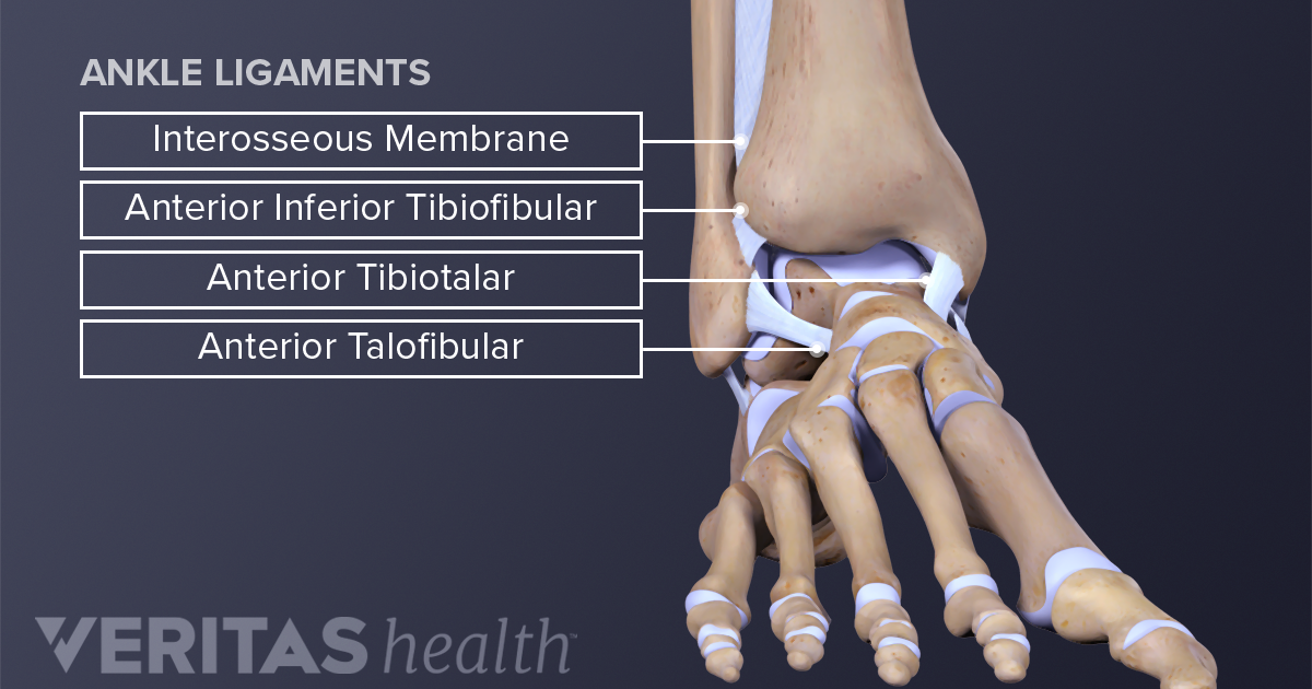
Ankle Anatomy Muscles And Ligaments from embed.widencdn.net Flexor tendon lacerations are classified into five zones 2, 15, 16. Jul 03, 2018 · in the calf region of the leg, the gastrocnemius muscle extends from the distal end of the femur through the calcaneal (achilles) tendon to the calcaneus of the heel. Jul 05, 2018 · the foot diagram has a complex structure made up of bones, ligaments, muscles, and tendons. As the damage progresses, the tendon can completely tear, sometimes when lifting a heavy object. (25 x 9.81) x 0.71 = 174nm (5.07 x 9.81) x 0.34 = 16.91nm = 174 + 16.91 = 190.91nm. Jul 01, 2021 · free body diagram for calculating deltoid force. If you would like to learn all the parts of the foot structure, you have come to the right place. In some cases, swelling occurs along with heat and redness, and there may be visible knots surrounding the joint.
Tendon reflexes, also referred to as myotatic reflexes, are variably affected by lower motor neuron lesions.
A zone 1 injury involves an fdp tendon laceration distal to the fds insertion. (25 x 9.81) x 0.71 = 174nm (5.07 x 9.81) x 0.34 = 16.91nm = 174 + 16.91 = 190.91nm. Hypoactive or absent reflexes are commonly observed in association with lower motor neuron lesions. A cut flexor tendon injury is a serious injury. Diagram illustrating tendonitis and tendon rupture symptoms can vary from aches or pains and local joint stiffness , to a burning that surrounds the whole joint around the inflamed tendon. If the complete motor neuron innervation to a muscle is lost, the efferent limb of a tendon reflex will fail. Understanding the structure of the foot is best done by looking at a foot diagram where the anatomy has been labeled. Gliding now medially shows the insertion of the subscapularis tendon. Problems after flexor tendon injury. The wound may be small, but the large forces carried by flexor tendons and the tendency for the repaired tendon to stick to the walls of the tunnel mean that despite a skilled repair and good hand therapy, many fingers do not regain full movement. In some cases, swelling occurs along with heat and redness, and there may be visible knots surrounding the joint. The subscapularis tendon lies approximately 3 to 5 cm under the surface. The fdp laceration is usually treated with fdp tendon advancement if the distal stump length is less than 1 cm or with tenorrhaphy if the distal stump length is greater than 1 cm 2, 17.