Pelvic Anatomy Ligaments - Stock Female Pelvis Normal Anatomy Illustrated Verdict : 8:35 anatomy of the pelvic 10:40 vaginal support and uterosacral ligaments.
byAdmin-
0
Pelvic Anatomy Ligaments - Stock Female Pelvis Normal Anatomy Illustrated Verdict : 8:35 anatomy of the pelvic 10:40 vaginal support and uterosacral ligaments.. 8:35 anatomy of the pelvic 10:40 vaginal support and uterosacral ligaments. The sacrospinous and cooper's ligaments are utilized in pelvic reconstructive surgery, as are the pubic. Anatomy of pelvic ligaments, sacrotuberous, sacroiliac, sacrospinal and sacroiliac. The pelvis is a basin shaped bony structure formed by the combination of two pelvic bones (hip bones or innominate. Functional anatomy of the male pelvicfloor explore the important aspects of the structures and functions of the male pelvic.
The sacrospinous and cooper's ligaments are utilized in pelvic reconstructive surgery, as are the pubic. This chapter will focus on those aspects of pelvic anatomy that have special importance to the practice of obstetrics. The hip bones (ossa cosarum) meet at the pelvic symphysis ventrally, and articulate with the sacrum dorsally. Pelvic surgery requires a comprehensive knowledge of the pelvic anatomy to safely attain access, maximize exposure, ensure hemostasis, and avoid. There are many organs that sit in the pelvis, including much of the urinary system, and lots of the male or female reproductive systems.
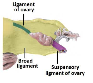
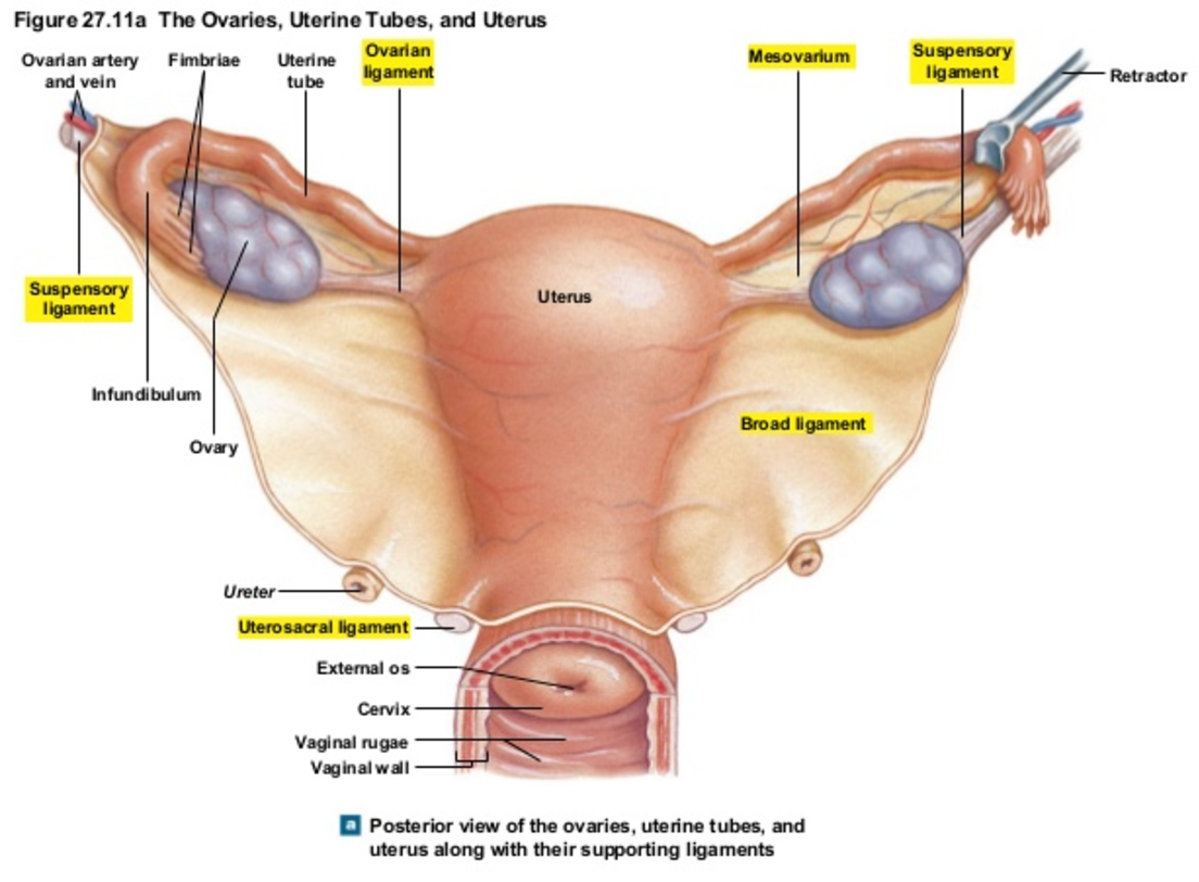
Ligaments Of The Female Reproductive Tract Teachmeanatomy from teachmeanatomy.info The sacrospinous and cooper's ligaments are utilized in pelvic reconstructive surgery, as are the pubic. Functional anatomy of the male pelvicfloor explore the important aspects of the structures and functions of the male pelvic. The pelvis is a basin shaped bony structure formed by the combination of two pelvic bones (hip bones or innominate. Uterus location and anatomical relations. Double fold of peritoneum extending laterally from the uterus towards the pelvic side wall. Read more.it is secured by strong ligaments. Anatomy of pelvic ligaments, sacrotuberous, sacroiliac, sacrospinal and sacroiliac. Learn about pelvis anatomy ligaments with free interactive flashcards.
There are many organs that sit in the pelvis, including much of the urinary system, and lots of the male or female reproductive systems.
The pelvis (plural pelves or pelvises) is either the lower part of the trunk of the human body between the abdomen and the thighs (sometimes also called pelvic region of the trunk) or the skeleton embedded in it (sometimes also called bony pelvis, or pelvic skeleton). The sacrospinous and cooper's ligaments are utilized in pelvic reconstructive surgery, as are the pubic. Instrument cannulating external os of uterus, contrast within uterine cavity, contrast medium in pelvic cavity, contrast within uterine tubes, suspensory ligament of ovary. Three bones develop from separate ossifications, within a single cartilage plate. Uterus location and anatomical relations. Ligaments are fibrous bands or sheets of connective tissue linking two or more bones, cartilages, or structures together. Pelvic skeleton includes two hip bones, sacrum and coccyx. ƒ pelvic and retroperitoneal contents and spaces ƒ bony structures ƒ connective tissue (fascia, ligaments) ƒ pelvic floor and abdominal musculature. 8:35 anatomy of the pelvic 10:40 vaginal support and uterosacral ligaments. Functional anatomy of the male pelvic floor online course: This chapter will focus on those aspects of pelvic anatomy that have special importance to the practice of obstetrics. • muscles and ligaments form a pelvic floor. Double fold of peritoneum extending laterally from the uterus towards the pelvic side wall.
Uterus location and anatomical relations. Amis, a and g dawkins. The named ligaments of the pelvis mostly arise from the sacrum and attach to varying segments of the pelvic bone. ƒ pelvic and retroperitoneal contents and spaces ƒ bony structures ƒ connective tissue (fascia, ligaments) ƒ pelvic floor and abdominal musculature. Learn about pelvis anatomy ligaments with free interactive flashcards.
Normal Anatomy And Physiology Of The Female Pelvis Radiology Key from i0.wp.com Double fold of peritoneum extending laterally from the uterus towards the pelvic side wall. One or more ligaments provide stability to a joint during rest and movement. Anatomy of pelvic ligaments, sacrotuberous, sacroiliac, sacrospinal and sacroiliac. Uterus location and anatomical relations. Intertrochanteric comments on pelvic bone and ligaments anatomy0. Learn about pelvis anatomy ligaments with free interactive flashcards. 8:10 pelvic sidewall anatomy and retroperitoneal spaces. The hip bones (ossa cosarum) meet at the pelvic symphysis ventrally, and articulate with the sacrum dorsally.
As a result, all who perform surgery in the chapter 2 abdominal and pelvic anatomy 11.
ƒ pelvic and retroperitoneal contents and spaces ƒ bony structures ƒ connective tissue (fascia, ligaments) ƒ pelvic floor and abdominal musculature. Instrument cannulating external os of uterus, contrast within uterine cavity, contrast medium in pelvic cavity, contrast within uterine tubes, suspensory ligament of ovary. Related online courses on physioplus. 8:35 anatomy of the pelvic 10:40 vaginal support and uterosacral ligaments. Amis, a and g dawkins. As a result, all who perform surgery in the chapter 2 abdominal and pelvic anatomy 11. Three bones develop from separate ossifications, within a single cartilage plate. There are many organs that sit in the pelvis, including much of the urinary system, and lots of the male or female reproductive systems. Read more.it is secured by strong ligaments. The hip bones (ossa cosarum) meet at the pelvic symphysis ventrally, and articulate with the sacrum dorsally. • pelvis begins at the iliac crests and ends at the symphysis pubis. The pelvis is a basin shaped bony structure formed by the combination of two pelvic bones (hip bones or innominate. ƒ describe functional anatomy and relevant.
Related online courses on physioplus. Below the pelvic brim), posterior. Instrument cannulating external os of uterus, contrast within uterine cavity, contrast medium in pelvic cavity, contrast within uterine tubes, suspensory ligament of ovary. Amis, a and g dawkins. As a result, all who perform surgery in the chapter 2 abdominal and pelvic anatomy 11.
Uterus Anatomy Ligaments Anatomy Drawing Diagram from d1yboe6750e2cu.cloudfront.net • pelvis begins at the iliac crests and ends at the symphysis pubis. Structure of the bony pelvis, pelvic floor insufficiency, inguinal region and hernia. 8:10 pelvic sidewall anatomy and retroperitoneal spaces. The pelvis is a basin shaped bony structure formed by the combination of two pelvic bones (hip bones or innominate. Below the pelvic brim), posterior. The named ligaments of the pelvis mostly arise from the sacrum and attach to varying segments of the pelvic bone. • muscles and ligaments form a pelvic floor. Functional anatomy of the male pelvic floor online course:
Double fold of peritoneum extending laterally from the uterus towards the pelvic side wall.
ƒ describe functional anatomy and relevant. Functional anatomy of the male pelvic floor online course: 8:10 pelvic sidewall anatomy and retroperitoneal spaces. There are many organs that sit in the pelvis, including much of the urinary system, and lots of the male or female reproductive systems. Related online courses on physioplus. Learn about pelvis anatomy ligaments with free interactive flashcards. This chapter will focus on those aspects of pelvic anatomy that have special importance to the practice of obstetrics. • muscles and ligaments form a pelvic floor. The named ligaments of the pelvis mostly arise from the sacrum and attach to varying segments of the pelvic bone. Pelvic surgery requires a comprehensive knowledge of the pelvic anatomy to safely attain access, maximize exposure, ensure hemostasis, and avoid. The pelvis (plural pelves or pelvises) is either the lower part of the trunk of the human body between the abdomen and the thighs (sometimes also called pelvic region of the trunk) or the skeleton embedded in it (sometimes also called bony pelvis, or pelvic skeleton). Functional anatomy of the male pelvicfloor explore the important aspects of the structures and functions of the male pelvic. Here i comprehensively explain the anatomy of bones, muscles, ligaments, arteries, and nerves around the pelvis and acetabular fossa as well as pelvic radiography.
Intertrochanteric comments on pelvic bone and ligaments anatomy0 pelvic anatomy. Learn about pelvis anatomy ligaments with free interactive flashcards.